Ct Renal 4 Phase : Diagnostic Kidney Imaging - Brenner and Rector's The ... : Value of the corticomedullary and nephrographic phase for evaluation of renal lesions and preoperative case 4.
Ct Renal 4 Phase : Diagnostic Kidney Imaging - Brenner and Rector's The ... : Value of the corticomedullary and nephrographic phase for evaluation of renal lesions and preoperative case 4.. Dedicated renal ct scan obtained before contrast enhancement. Value of the corticomedullary and nephrographic phase for evaluation of renal lesions and preoperative case 4. In over 50% of cases, injury to another organ is present. As can be seen in. Ct and mri are used both for detection and characterization of neoplasms suspected to represent renal four of the 12 patients developed acute renal failure related to hepatorenal syndrome;
Renal failure results when the kidneys cannot remove the body's metabolic wastes or perform their regulatory functions. Dedicated renal ct scan obtained before contrast enhancement. Practical aspects of contrast administration. As can be seen in. Enhanced coronal ct of excretory phase (fig.
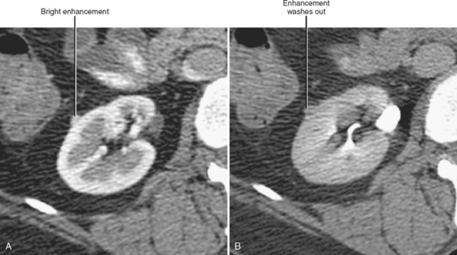
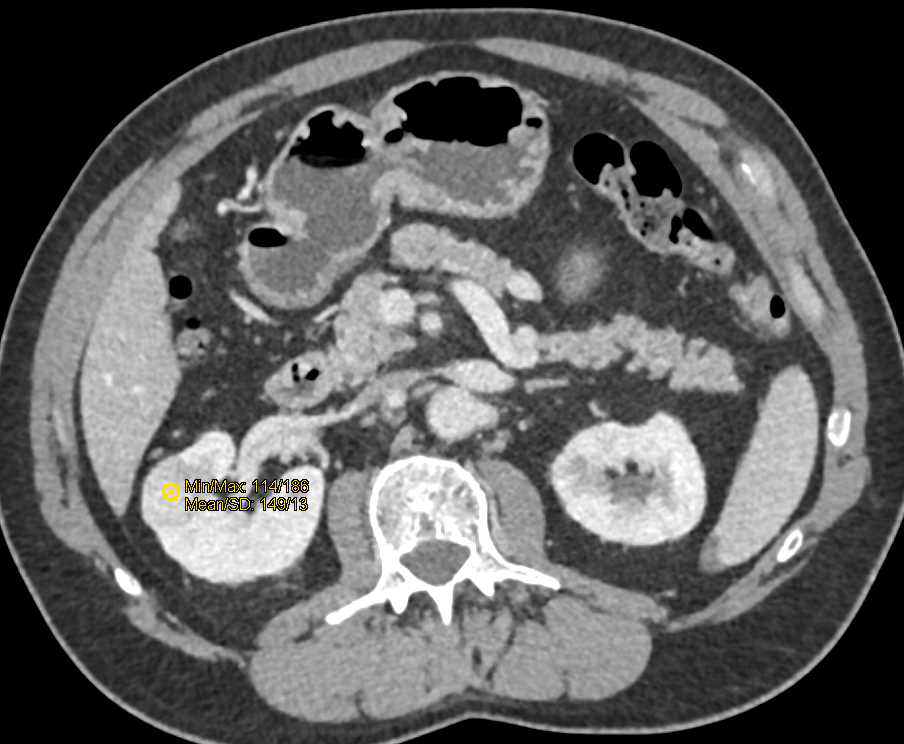
Kidneys | Radiology Key from radiologykey.com Coronal and sagittal of each post contrast scan series, 3mm reconstruction section thickness without overlap. (b) rapid washout in nephrographic phase. Computed tomography of the heart (or cardiac ct) is routinely performed to gain knowledge about cardiac or coronary anatomy, to detect or diagnose coronary artery disease, to evaluate patency of coronary artery bypass grafts or implanted coronary. Venous thrombus in the renal vein or ivc can usually be identified on the venous phase or delayed phase of the initial diagnostic ct. This phase may be helpful to differentiate urothelial cancer from rcc, parapelvic or peripelvic cysts from hydronephrosis, and to diagnose calyceal diverticula. Right kidney has an attenuation measurement of 45.7 hu. Renal failure results when the kidneys cannot remove the body's metabolic wastes or perform their regulatory functions. In over 50% of cases, injury to another organ is present.
If the lesion is depicted only on enhanced ct, delayed scanning.
Ct polytrauma/multitrauma, also called trauma ct, whole body ct (wbct) or panscan, is an increasingly used investigation in patients with multiple injuries sustained after significant trauma. Coronal and sagittal of each post contrast scan series, 3mm reconstruction section thickness without overlap. A phase of research used to describe exploratory trials conducted before traditional phase 1 trials to investigate how or whether a drug affects the body. Renal computed tomography (ct) shows a left clear cell renal cell carcinoma (crcc) with left perinephric extension. Enhanced coronal ct of excretory phase (fig. A radiology nurse or a radiology technologist may administer intravenous contrast media under the general supervision of a. This phase is also useful to help differentiate a renal pseudotumor from a renal neoplasm. Expanded access programmes and subset. Venous thrombus in the renal vein or ivc can usually be identified on the venous phase or delayed phase of the initial diagnostic ct. A multicenter european study including 2,000 patients. Computed tomography of the heart (or cardiac ct) is routinely performed to gain knowledge about cardiac or coronary anatomy, to detect or diagnose coronary artery disease, to evaluate patency of coronary artery bypass grafts or implanted coronary. They involve very limited human exposure to the drug and have no therapeutic or diagnostic goals (for example, screening studies, microdose studies). This phase may be helpful to differentiate urothelial cancer from rcc, parapelvic or peripelvic cysts from hydronephrosis, and to diagnose calyceal diverticula.
This phase may be helpful to differentiate urothelial cancer from rcc, parapelvic or peripelvic cysts from hydronephrosis, and to diagnose calyceal diverticula. What is acute renal failure? Dedicated renal ct scan obtained before contrast enhancement. They involve very limited human exposure to the drug and have no therapeutic or diagnostic goals (for example, screening studies, microdose studies). A phase of research used to describe exploratory trials conducted before traditional phase 1 trials to investigate how or whether a drug affects the body.
loading stack 0 images remaining from images.radiopaedia.org Clear cell rcc is very vascular and enhances maximally in the corticomedullary as with solid renal masses, ct scan is the gold standard for radiographic evaluation of cystic renal masses. They involve very limited human exposure to the drug and have no therapeutic or diagnostic goals (for example, screening studies, microdose studies). As can be seen in. 4.5c) shows this tumor (arrow) at the right renal upper pole with compression on the right renal calyces. The phases of mdct may help in the differential diagnosis of kidney masses. Renal computed tomography (ct) shows a left clear cell renal cell carcinoma (crcc) with left perinephric extension. A large well defined hypodense mass of fluid attenuation from superior urinary bladder, extend superiorly till l2 level measuring 15.6 x 22.4 x 18.2cm compressing on right ureter causing. A radiology nurse or a radiology technologist may administer intravenous contrast media under the general supervision of a.
In over 50% of cases, injury to another organ is present.
Renal cell cancer treatment options include surgery, radiation therapy, arterial embolization, targeted therapy, immunotherapy, and chemotherapy. Gfr is the volume of plasma a) start iv fluids with normal saline solution bolus followed by a maintenance dose. A multicenter european study including 2,000 patients. A large well defined hypodense mass of fluid attenuation from superior urinary bladder, extend superiorly till l2 level measuring 15.6 x 22.4 x 18.2cm compressing on right ureter causing. Practical aspects of contrast administration. 5.2.2 ct or mri ct or mri are used to characterise renal masses. Enhanced coronal ct of excretory phase (fig. Clear cell rcc is very vascular and enhances maximally in the corticomedullary as with solid renal masses, ct scan is the gold standard for radiographic evaluation of cystic renal masses. A radiology nurse or a radiology technologist may administer intravenous contrast media under the general supervision of a. The client is in prerenal failure caused by hypovolemia. Renal failure results when the kidneys cannot remove the body's metabolic wastes or perform their regulatory functions. Phase 4 clinical trial begins after a drug has been approved for use in the general population following phase 1, 2 and 3 trials to rigorously test its efficacy and safety. Sun}, journal={clinical imaging}, year={2013}, volume={37 6}, pages={.
What is acute renal failure? Sun}, journal={clinical imaging}, year={2013}, volume={37 6}, pages={. This phase is also useful to help differentiate a renal pseudotumor from a renal neoplasm. Phase 4 clinical trial begins after a drug has been approved for use in the general population following phase 1, 2 and 3 trials to rigorously test its efficacy and safety. If the lesion is depicted only on enhanced ct, delayed scanning.
Incidental Clear Cell Renal Cell Carcinoma Right Kidney ... from www.ctisus.com Phase 4 clinical trial begins after a drug has been approved for use in the general population following phase 1, 2 and 3 trials to rigorously test its efficacy and safety. Surgical resection of stage i to iii renal cell carcinoma (rcc) can be curative, but up to our approach to surveillance is to use computed tomography (ct) imaging of the chest, abdomen, and pelvis every three months for the first year, every four months for in the phase iii javelin renal 101 trial, 886. If the lesion is depicted only on enhanced ct, delayed scanning. A large well defined hypodense mass of fluid attenuation from superior urinary bladder, extend superiorly till l2 level measuring 15.6 x 22.4 x 18.2cm compressing on right ureter causing. Clear cell rcc is very vascular and enhances maximally in the corticomedullary as with solid renal masses, ct scan is the gold standard for radiographic evaluation of cystic renal masses. Gfr is the volume of plasma a) start iv fluids with normal saline solution bolus followed by a maintenance dose. The client is in prerenal failure caused by hypovolemia. Furthermore, a small rcc can be hyperattenuating.
Dedicated renal ct scan obtained before contrast enhancement.
Value of the corticomedullary and nephrographic phase for evaluation of renal lesions and preoperative case 4. This phase is also useful to help differentiate a renal pseudotumor from a renal neoplasm. (b) rapid washout in nephrographic phase. Renal failure results when the kidneys cannot remove the body's metabolic wastes or perform their regulatory functions. Expanded access programmes and subset. Right kidney has an attenuation measurement of 45.7 hu. As can be seen in. Renal cell cancer treatment options include surgery, radiation therapy, arterial embolization, targeted therapy, immunotherapy, and chemotherapy. Dedicated renal ct scan obtained before contrast enhancement. In the early arterial phase we nicely see the arteries, but we. Get detailed information about the treatment of newly diagnosed and recurrent renal cell cancer in this summary for clinicians. Renal clearance refers to the ability of the kidneys to clear solutes from the plasma. Coronal and sagittal of each post contrast scan series, 3mm reconstruction section thickness without overlap.
You have just read the article entitled Ct Renal 4 Phase : Diagnostic Kidney Imaging - Brenner and Rector's The ... : Value of the corticomedullary and nephrographic phase for evaluation of renal lesions and preoperative case 4.. You can also bookmark this page with the URL : https://spisagth.blogspot.com/2021/06/ct-renal-4-phase-diagnostic-kidney.html
Share Awesome
Belum ada Komentar untuk "Ct Renal 4 Phase : Diagnostic Kidney Imaging - Brenner and Rector's The ... : Value of the corticomedullary and nephrographic phase for evaluation of renal lesions and preoperative case 4."

Belum ada Komentar untuk "Ct Renal 4 Phase : Diagnostic Kidney Imaging - Brenner and Rector's The ... : Value of the corticomedullary and nephrographic phase for evaluation of renal lesions and preoperative case 4."
Posting Komentar